

See how the DormoVision X™ Home PSG platform brings objective RBD assessment into the home, enabling detection and quantitative characterization of REM sleep without atonia (RSWA) beyond the traditional sleep laboratory.
Introduction
Rapid eye movement (REM) sleep behavior disorder (RBD) is a parasomnia characterized by dream-enactment behaviors associated with loss of the physiological muscle atonia normally present during REM sleep. An essential objective component of RBD diagnosis is the polysomnographic demonstration of REM sleep without atonia (RSWA), manifested by excessive muscle activity during REM sleep (Cesari et al., 2022; Howell et al., 2023). RBD is of particular clinical importance as a robust and highly specific predictor of neurodegeneration, with over 90% of individuals phenoconverting to Parkinson’s disease or other α-synucleinopathies within 14 years (Galbiati et al., 2019).
Quantitative analysis of REM EMG activity provides an objective measure of RSWA by differentiating sustained tonic activity from brief phasic bursts. Clinically validated thresholds have been established for quantifying abnormal REM EMG activity using chin and limb EMG, individually and in combination (Frauscher et al., 2012). This quantitative approach, however, has traditionally relied on laboratory-based polysomnography.
The DormoVision X™ Platform bridges this gap by bringing comprehensive PSG capabilities, including EEG, EOG, chin and leg EMG, into the home environment, providing the physiological signals required to identify REM sleep and quantitatively assess excessive RSWA outside the conventional sleep laboratory. Here, we present a single-patient case demonstrating the detection and quantitative characterization of RSWA using the DormoVisionX™ Type-2 Home PSG in a patient with clinically established RBD. This case demonstrates the potential of comprehensive home PSG to extend objective RSWA assessment beyond the sleep laboratory, providing a more accessible approach for evaluating and longitudinally monitoring REM-related abnormalities associated with neurodegenerative processes.
Methods
Patient and sleep recording
A 72-year-old woman with clinically established RBD underwent overnight Type-2 polysomnography. RBD was supported by a previous in-laboratory PSG and a clinical history of increased motor activity during sleep. The REM Sleep Behavior Disorder Screening Questionnaire (RBDSQ) score was 7/10.
The overnight recording was performed using the DormoVision X™ Type-2 Home PSG and included EEG, EOG, chin and leg EMG, airflow, SpO₂, PPG-derived cardiovascular measures, snoring, and body position.
RSWA detection and quantification
Sleep stages, respiratory events, and arousals were scored according to AASM guidelines. RSWA was quantified separately from the chin and leg EMG channels. Motor activity during REM sleep was classified as phasic, tonic, or any RSWA, following established SINBAR-based methodology (Frauscher et al., 2012). Each 30-s REM epoch was divided into ten 3-s mini-epochs. Phasic activity was defined as discrete EMG bursts lasting 0.1–5.0 s with an amplitude exceeding twice the background EMG activity. Tonic activity was identified at the 30-s epoch level when sustained elevation in EMG activity occupied >50% of the epoch. “Any” RSWA represented REM segments containing phasic, tonic, or mixed EMG activity. Periods affected by artifacts or EMG activity associated with respiratory events were excluded from the RSWA assessment.
Phasic and any RSWA indices were calculated as the percentage of valid 3-s REM mini-epochs classified as positive for the respective activity, whereas the tonic RSWA index was calculated as the percentage of valid 30-s REM epochs classified as tonic-positive. RSWA quantification was performed automatically by the DormoVisionX™ Platform, with the resulting indices incorporated into the final sleep report.
Results
The recording included 7 h 41 min of total sleep time, with 121 min spent in REM sleep, providing sufficient REM data for quantitative RSWA assessment. The patient had an RBDSQ score of 7/10, supporting the presence of clinical features associated with RBD. The patient also exhibited mild obstructive sleep apnea, with an apnea–hypopnea index (AHI) of 7.8 events/h using the 4% desaturation criterion. Additional clinical and sleep parameters are summarized in Table 1.
Both chin and leg EMG demonstrated increased motor activity during REM sleep, including tonic activity and phasic bursts distributed across REM episodes (Figure 1A–B). However, chin EMG showed greater sensitivity for detecting RSWA, particularly tonic activity, with a tonic RSWA index of 33.5% for chin EMG compared with 2.1% for leg EMG. This pattern is consistent with previous evidence supporting the high diagnostic sensitivity of mentalis EMG for RSWA detection and its utility for quantitative assessment of REM motor abnormalities (Frauscher et al., 2012; Lanir-Azaria et al., 2026).
Figure 1C presents the quantitative RSWA indices derived from chin EMG. The phasic, tonic, and any RSWA indices all exceeded their respective clinically validated thresholds, demonstrating excessive RSWA consistent with the patient’s established RBD diagnosis.
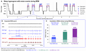
Figure 1—Detection and quantification of REM sleep without atonia (RSWA).
(A) Full-night sleep hypnogram showing sleep-stage distribution and motor events occurring during REM sleep. Phasic and tonic events detected in the chin and leg EMG channels are indicated by the corresponding markers. The highlighted segment within the second REM episode indicates the 1-min interval selected for detailed visualization. (B) Representative 1-min REM segment displaying EEG, EOG, chin EMG, and leg EMG signals. Chin EMG demonstrates sustained tonic activity followed by multiple phasic bursts, illustrating both tonic and phasic RSWA. (C) Quantitative chin EMG RSWA indices for phasic, tonic, and any RSWA. Dashed lines indicate the respective upper limits of normal (10.6%, 8.7%, and 14.5%); values above each threshold are shown in the darker shade. The measured phasic, tonic, and any RSWA indices were 14.2%, 33.5%, and 43.9%, respectively, all exceeding their corresponding thresholds. Thresholds were based on previously established quantitative criteria for mentalis EMG RSWA (Frauscher et al., 2012).
| Patient characteristics | |||
| Parameter | Value | Parameter | Value |
|---|---|---|---|
| Age | 72 years | Sex | Female |
| BMI | 27.9 kg/m² | RBDSQ | 7/10 |
| Sleep parameters & architecture | |||
| Parameter | Value | Parameter | Value |
|---|---|---|---|
| Total recording time | 7 h 47 min | Total sleep time | 7 h 41 min |
| Sleep efficiency | 98.5% | Sleep latency | 4.0 min |
| REM latency | 110.0 min | WASO | 2.9 min |
| N1 sleep | 3.5 min (1%) | N2 sleep | 265.0 min (57%) |
| N3 sleep | 71.5 min (16%) | REM sleep | 121.0 min (26%) |
| Arousal index | 5.5 events/h | ||
| RSWA indices | ||
| Parameter | Chin EMG | Leg EMG |
|---|---|---|
| Phasic RSWA index | 14.2% | 6.6% |
| Tonic RSWA index | 33.5% | 2.1% |
| Any RSWA index | 43.9% | 8.7% |
| Respiratory parameters | ||
| Parameter | 3% criterion | 4% criterion |
|---|---|---|
| AHI | 19.1 events/h | 7.8 events/h |
| REM AHI | 24.8 events/h | 11.9 events/h |
| ODI | 15.9 events/h | 11.1 events/h |
| Minimum SpO2 | 82% | |
Conclusion
This case demonstrates the feasibility of detecting and quantitatively characterizing RSWA using comprehensive Type-2 home PSG. The DormoVision X™ Platform captured both phasic and tonic REM-related motor activity and provided quantitative RSWA indices consistent with the patient’s established RBD diagnosis. These findings support the potential of comprehensive home PSG to extend objective RBD assessment beyond the conventional sleep laboratory, enabling more accessible and longitudinal evaluation of REM-related motor abnormalities.
References
Cesari, M., Heidbreeder, A., St. Louis, E. K., Sixel-Döring, F., Bliwise, D. L., Baldelli, L., Bes, F., Fantini, M. L., Iranzo, A., Knudsen-Heier, S., Mayer, G., McCarter, S. J., Nepozitek, J., Plazzi, G., Provini, F., Santamaria, J., Sunwoo, J. S., Videnovic, A., & Högl, B. (2022). Video-polysomnography procedures for diagnosis of rapid eye movement sleep behavior disorder (RBD) and the identification of its prodromal stages: Guidelines from the International RBD Study Group. Sleep, 45(3), zsab257.
Frauscher, B., Iranzo, A., Gaig, C., Gschliesser, V., Guaita, M., Raffelseder, V., Ehrmann, L., Sola, N., Salamero, M., Tolosa, E., Poewe, W., Santamaria, J., & SINBAR (Sleep Innsbruck Barcelona) Group. (2012). Normative EMG values during REM sleep for the diagnosis of REM sleep behavior disorder. Sleep, 35(6), 835–847.
Galbiati, A., Verga, L., Giora, E., Zucconi, M., & Ferini-Strambi, L. (2019). The risk of neurodegeneration in REM sleep behavior disorder: A systematic review and meta-analysis of longitudinal studies. Sleep Medicine Reviews, 43, 37–46.
Howell, M., Avidan, A. Y., Foldvary-Schaefer, N., Malkani, R. G., During, E. H., Roland, J. P., McCarter, S. J., Zak, R. S., Carandang, G., Kazmi, U., & Ramar, K. (2023). Management of REM sleep behavior disorder: An American Academy of Sleep Medicine clinical practice guideline. Journal of Clinical Sleep Medicine, 19(4), 759–768.
Lanir-Azaria, S., Nir, Y., Tauman, R., Zitser, J., & Giladi, N. (2026). Beyond RBD: Covert REM sleep abnormalities in Parkinson’s disease. npj Parkinson’s Disease, 12, 90.
Regulatory information
DormoVision X™ is the commercial platform utilizing the FDA-cleared Dormotech NLab™ and Vlab™ physiological data recording systems (510(k) K242290 and 510(k) K230148). The platform is indicated for prescription use by or on the order of a licensed physician for the acquisition and display of comprehensive physiological sleep parameters in both supervised (clinical/laboratory) and unsupervised (home) environments for patients greater than 6 years of age. Automated scoring and analytical features function strictly as data visualization aids and do not supersede manual review or clinical interpretation by a qualified sleep technologist or physician.